


Copyright 2016 Biomedical Microdevices Laboratory
Current Research
Representative publications:
[1] PT McCarthy, KJ Otto, and MP Rao. Biomed Microdevices 13(3):503-515, 2011.
[2] PT McCarthy, MP Rao, and KJ Otto. J Neural Engr 8(4):046007 (9pp), 2011.
[1] PT McCarthy, KJ Otto, and MP Rao. Biomed Microdevices 13(3):503-515, 2011.
[2] PT McCarthy, MP Rao, and KJ Otto. J Neural Engr 8(4):046007 (9pp), 2011.
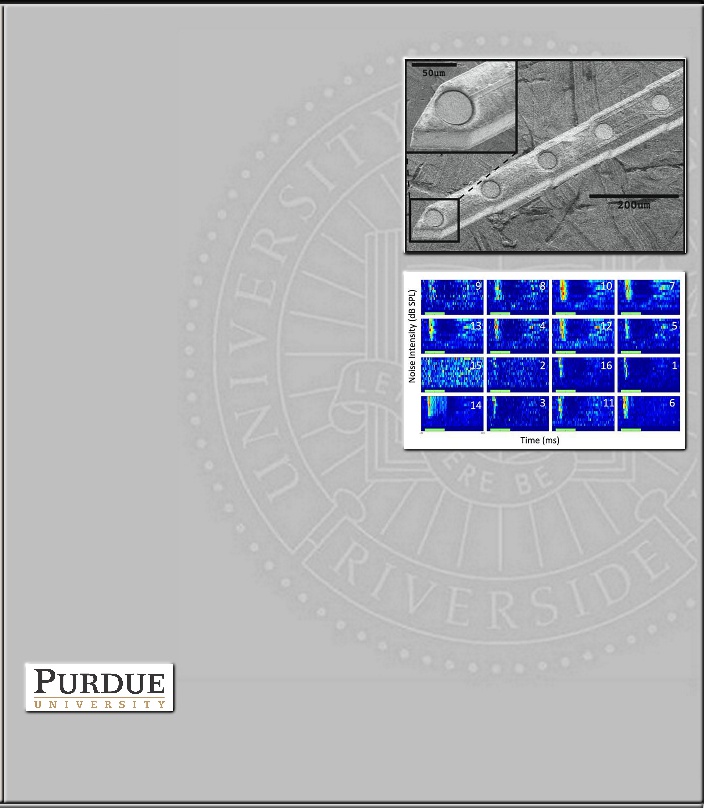
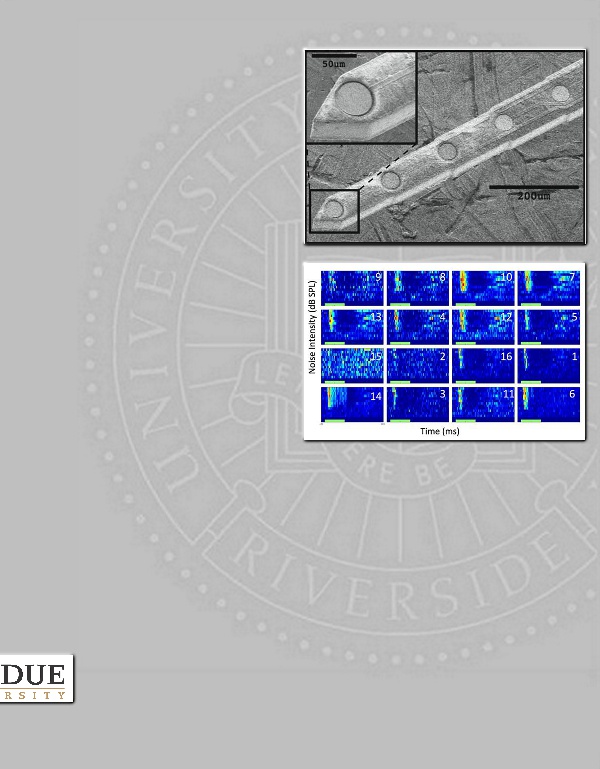
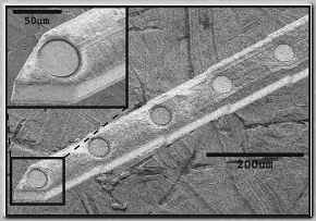
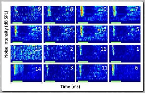
Top: Scanning electron micrograph of
Ti-based penetrating microelectrode [1].
Bottom: Simultaneously-recorded,
noise-evoked, peri-stimulus time
histograms from the auditory cortex (top
2 rows) and thalamus (bottom 2 rows) of
an anesthetized rat. The green bar along
the x-axis reflects the timing of the
broadband acoustic noise stimulus [2].
Penetrating microelectrodes
Research in this thrust focuses on addressing fundamental reliability limitations that may ultimately constrain the clinical translation of penetrating microelectrodes used for neural prosthetic interfaces.
By providing means for directly interacting with neural tissues, penetrating microelectrodes have shown promise for restoring neurological functions lost to disease, stroke, or injury. However, the intrinsic brittleness of silicon, the material most commonly used for the manufacture of such devices, creates non-negligible probability for fracture-based fragmentation within the brain. This therefore motivates the development of means for mitigating this hazard, given the potential severity and resulting adverse impact this could have on future clinical viability.
The Ti-based microelectrodes shown here seek to address this limitation. Thus far, we have demonstrated that these devices: a) provide potential for enhanced safety, due to their graceful, plasticity-based failure mode [1]; b) posses sufficient stiffness for reliable cortical penetration [1]; c) provide recording performance comparable to that of commercially-available devices [1]; and d) allow, for the first time, simultaneous recording of multi-unit data and isolated action potentials in auditory cortex and thalamus [2].
Collaborators:
KJ Otto, BME, Purdue University
Sponsors:
Showalter Research Trust (PI: Rao)
Showalter Research Trust (PI: Rao)