


Copyright 2016 Biomedical Microdevices Laboratory
Current Research
Representative publications:
[1] J Lu, MP Rao, NC MacDonald, D Khang, & TJ Webster. Acta Biomater 4(1):192-201, 2008.
[2] P Vandrangi, SC Gott, VGJ Rodgers, & MP Rao. MMB 2013.
[3] P Vandrangi, SC Gott, R Kozaka, MP Rao, & VGJ Rodgers. Circulation 128:A18077, 2013.
[4] MP Rao, J Lu, HP Aguilar, NC MacDonald, & TJ Webster. Hilton Head 2008.
[5] 3. P Vandrangi, SC Gott, R Kozaka, VGJ Rodgers, & MP Rao. PLoS ONE 9(10):e111465, 2014.
[6] SC Gott, BA Jabola, & MP Rao. J Micromech Microeng 25(8):085016, 2015.
Vascular intervention
Research in this thrust focuses on developing deeper understanding of how nanoscale surface topography affects vascular cell response, and applying this understanding towards rational design of stents and other devices to elicit therapeutically desirable outcomes.
Thus far, our in vitro studies on planar substrates have shown that rationally-designed surface nanopatterning can: a) enhance endothelial cell (EC) adhesion and proliferation [1-3]; b) promote elongated EC morphology reminiscent of the native endothelium [1-3]; c) preferentially enhance EC adhesion and proliferation over smooth muscle cells under competitive co-culture conditions [4]; and d) promote enhanced atheroprotective factor expression in ECs [3,5]. Collectively, these results suggest potential for a new paradigm where rationally-designed nanopatterning provides a physical means for complementing (or replacing) current pharmacological intervention schemes for mitigating adverse physiological responses to stenting (i.e. restenosis & late stent thrombosis).
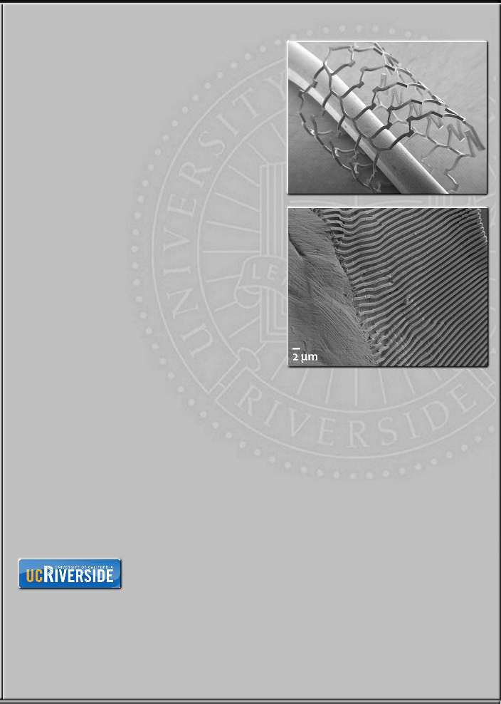
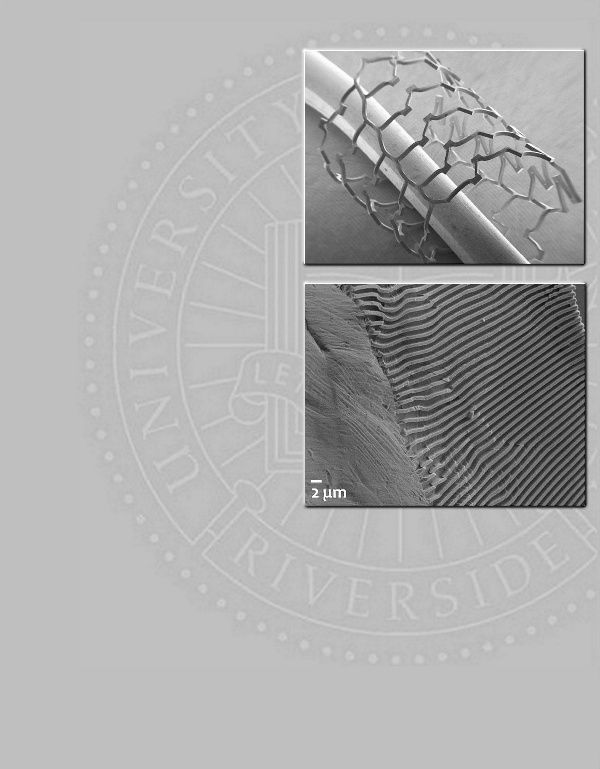
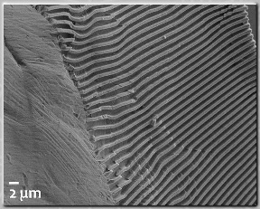
In parallel, we have been developing means for evaluating cellular response to surface nanopatterning in more physiologically-relevant contexts, i.e. those that provide exposure to the complex multicellular milieu, flow-induced shear, and tissue-device interactions present in vivo. The balloon-deployable nanopatterned stents shown here represent the first attempt to develop a device platform that will eventually provide such capability, and do so in a manner consistent with conventional vascular stents [6].
Collaborators:
VGJ Rodgers, BIEN, UCR
K Ghosh, BIEN, UCR
G Xu, ME, UCR
Research in this thrust focuses on developing deeper understanding of how nanoscale surface topography affects vascular cell response, and applying this understanding towards rational design of stents and other devices to elicit therapeutically desirable outcomes.
Thus far, our in vitro studies on planar substrates have shown that rationally-designed surface nanopatterning can: a) enhance endothelial cell (EC) adhesion and proliferation [1-3]; b) promote elongated EC morphology reminiscent of the native endothelium [1-3]; c) preferentially enhance EC adhesion and proliferation over smooth muscle cells under competitive co-culture conditions [4]; and d) promote enhanced atheroprotective factor expression in ECs [3,5]. Collectively, these results suggest potential for a new paradigm where rationally-designed nanopatterning provides a physical means for complementing (or replacing) current pharmacological intervention schemes for mitigating adverse physiological responses to stenting (i.e. restenosis & late stent thrombosis).
In parallel, we have been developing means for evaluating cellular response to surface nanopatterning in more physiologically-relevant contexts, i.e. those that provide exposure to the complex multicellular milieu, flow-induced shear, and tissue-device interactions present in vivo. The balloon-deployable nanopatterned stents shown here represent the first attempt to develop a device platform that will eventually provide such capability, and do so in a manner consistent with conventional vascular stents [6].
Collaborators:
VGJ Rodgers, BIEN, UCR
K Ghosh, BIEN, UCR
G Xu, ME, UCR
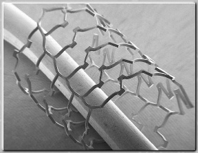
Top: Scanning electron micrograph of
nanopatterned Ti stent (deployed).
Bottom: Surface of a nanopatterned
stent with 0.75 µm groove width grating
in heavily deformed region [6].
Sponsors:
UCR Collaborative Seed Grant Program (PI: Rao)
UCR Collaborative Seed Grant Program (PI: Rao)